MIT News
April 7, 2025
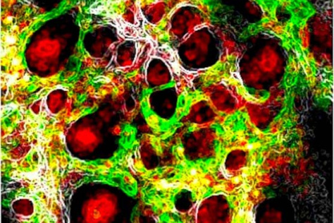
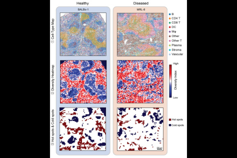
By treating diseased tissue as an ecosystem, Alex Shalek and team codeveloped MESA, a tool that reveals hidden interactions between cancer and immune cells. Their research, published in Nature Genetics, shows that when applied to a diverse range of cancer tissue types, including colorectal and liver cancer, MESA uncovered critical hotspots of cellular activity, offering new insights into disease progression.