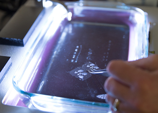
A formalin-fixed paraffin-embedded tissue sample
Going back decades, biopsies, tumor sections, and other tissue samples from cancer patients have been preserved by a two-step process: first the sample is fixed in formaldehyde, or formalin, to preserve proteins and vital structures within the tissue. Then, it is embedded in a block of paraffin wax, which makes it easier to slice samples into the sizes required for mounting on a microscope slide. These formalin-fixed paraffin-embedded (FFPE) tissues serve as a rich repository of clinical samples for discovery and translational research. However, FFPE samples often contain only small amounts of tissue, making them unsuitable for certain types of analysis.
Such has been the case in the White Laboratory, which has developed techniques for analyzing cell signaling processes and identifying potential cancer therapy targets and drug combinations. A molecular signaling process of particular interest to the group is tyrosine phosphorylation, where in-depth studies and quantification of changes in kinase signaling networks in various disease and treatment contexts have previously required large amounts of tissue contained in frozen patient samples, a less common form of preservation. Their analysis, which combines mass spectrometry with computational modeling, is powerful, but has not been compatible with FFPE samples—until now.
A recent Cancer Research paper describes an updated method developed by White lab researchers, and tested with collaborators at the Mayo Clinic, that now allows them to apply their highly sensitive, quantitative analysis of tyrosine phosphorylation signaling networks to FFPE tissues, and to obtain the same valuable data as from frozen samples. Using the new technique to analyze FFPE samples of breast and lung tumors, the team was able to identify patient-specific cancer-driving signaling molecules, indicating potential targeted therapies for each patient. These findings suggest the ability of this technique to provide direct translational insight from tyrosine phosphorylation analysis of small amounts of FFPE tumor tissue specimens, and open huge repositories of human samples to further study.